
An interesting topic came up in an online dental forum around what to do when the dental professional notices an indication on a panoramic radiograph that resembles calcification in the carotid artery.
Based on this, we wanted to provide a few resources that may be useful for dental staff to be aware of this indication along with potential implications.
Make sure you stay updated on the latest in the dental industry by subscribing
Carotid artery calcifications (CAC) are hard, calcified deposits that form in the carotid arteries, which are the major blood vessels in the neck. There are two carotid arteries, one on each side of the neck: the right and left common carotid arteries. Each common carotid artery branches into the internal carotid artery, which supplies blood to the brain, and the external carotid artery, which supplies blood to the face and neck.
These calcifications can indicate a higher risk of cardiovascular diseases like stroke and heart attack. Detecting these early can be vital for preventing serious health issues.
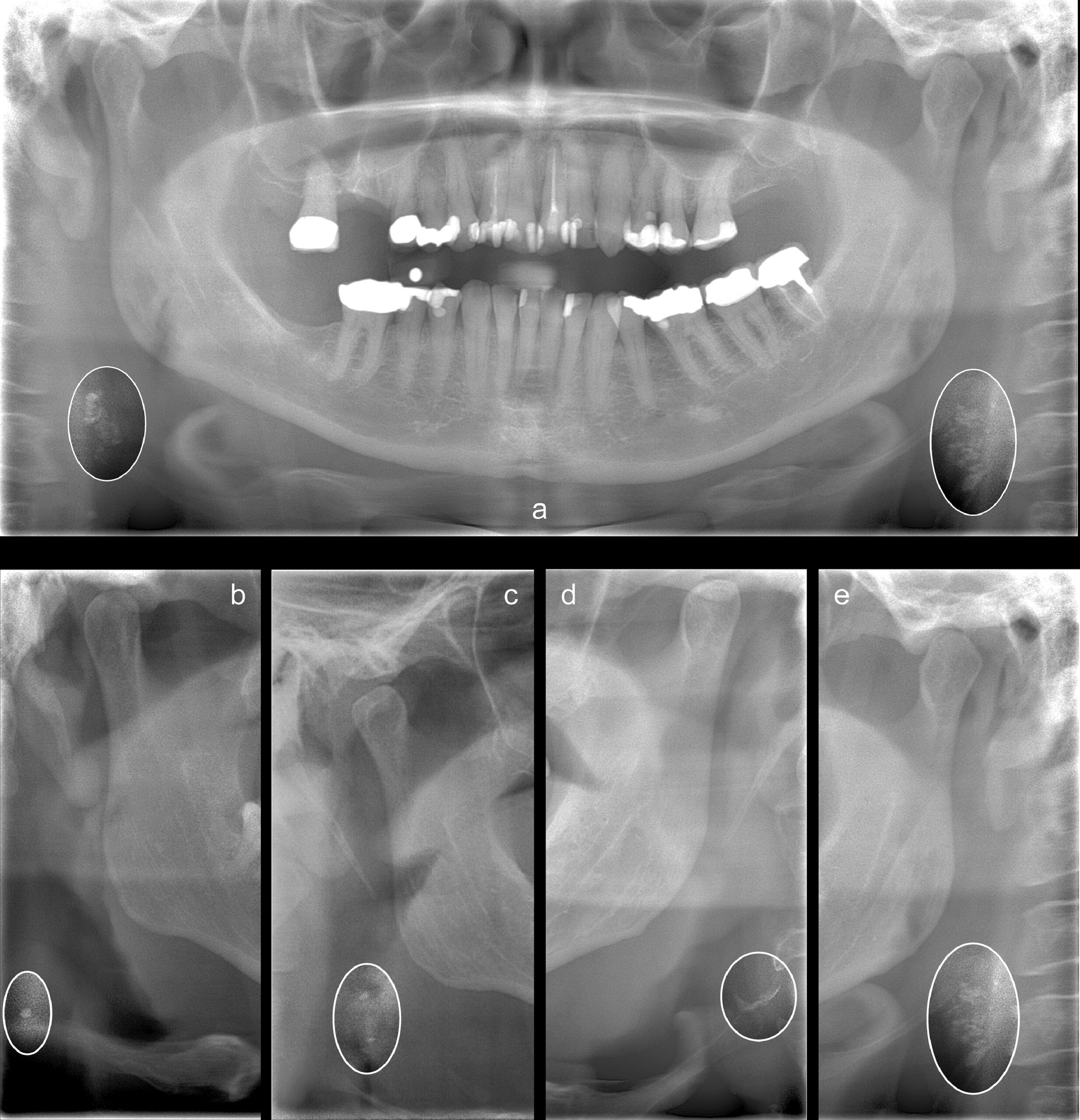
Dental panoramic radiographs can also show the carotid arteries. When dentists review these radiographs, they might notice radiopaque nodular lesions, which can indicate CAC. According to Friedlander et al. [1], these lesions appear separate from the hyoid bone and are adjacent to the cervical vertebrae, usually between the C3 and C4 vertebrae or below. This is close to the location where the carotid artery bifurcates.
Research Findings
Studies such as Magnus Bladh et. al [2] have shown a strong correlation between the presence of CAC on panoramic radiographs and more advanced carotid atherosclerosis detected by ultrasound (US). Ultrasound is a well-established method for detecting soft tissue changes and plaques in the arteries, which are significant markers for cardiovascular disease. The findings suggest that if CAC is detected on a panoramic radiograph, there is a high probability that more severe atherosclerotic changes will be found upon further examination with ultrasound.
Here are a few sample radiographs from this study:
Understanding the panoramic x-ray projection
Because all panoramic radiographs are projections, it’s important to be aware that the farther from the focal trough the anatomy is, the more distorted it may appear. In this situation, distortion is not as important because it’s more a matter of flagging the indication. However, if you are using a panoramic x-ray machine that has the ability to capture multiple focal troughs, then it is possible to gather additional views from the single scan. In other words, these panoramic x-ray machines allow the operator to extract more information from the scan by evaluating focal planes that may not be as ideal for the dentition, but more ideal to view the carotid artery.
Advantages and Limitations
The ability to detect CAC on panoramic radiographs can be particularly useful in both general and specialized dentistry. Dentists who identify these calcifications can recommend that patients seek medical care for further evaluation and preventive treatment. This can help reduce the risk of CVD-related issues.
However, it’s important to note that not all carotid plaques are calcified, and sometimes the plaques may be located outside the area captured by the X-ray. Therefore, a negative finding on a panoramic radiograph does not necessarily mean that the patient has no vascular issues.
Despite some limitations, the high specificity of CAC detected on panoramic radiographs, coupled with their correlation to ultrasound findings, underscores their importance. While ultrasound can visualize the soft tissue components of plaques that X-rays cannot, panoramic X-rays excel at detecting even small calcifications [2].
In some cases, calcifications may not be visible on panoramic radiographs if they are situated below the imaging area. Yet, when CACs are detected, particularly when they are bilateral and outline the vessel, it strongly suggests significant vascular changes that require medical attention [2].
Conclusion
In summary, panoramic radiographs are a valuable tool in dental practice not only for assessing dental health but also for identifying potential cardiovascular risks. Dentists who recognize CACs on these radiographs can play a crucial role in early detection and prevention of cardiovascular disease. By recommending further medical evaluation for patients with detected CACs, dentists can contribute to improved cardiovascular health outcomes.
While panoramic X-rays should not replace traditional cardiovascular screenings, they offer an additional layer of detection that can be lifesaving. For patients, this means that routine dental exams could also provide important insights into their overall health, highlighting the interconnected nature of dental and medical care.
- AH Friedlander, NR Garrett, EE Chin, JD. Baker; Ultrasonographic confirmation of carotid artery atheromas diagnosed via panoramic radiography; J Am Dent Assoc, 136 (2005), pp. 635-640
- Magnus Bladh, DDS,a Nils Gustafsson, DDS, PhD, et.al; Defined shapes of carotid artery calcifications on panoramic radiographs correlate with specific signs of cardiovascular disease on ultrasound examination; Oral Surgery, Oral Medicine, Oral Pathology and Oral Radiology Volume 137, Issue 4, April 2024, Pages 408-420
Make sure you stay updated on the latest in the dental industry by subscribing