If my X-rays are coming out too light or too dark, what can I tell my staff to do differently?
As much as traditional intraoral radiography has become a rudimentary part of the office standard operating procedure, it can be easy to forget that there are a ton of variables that can affect the diagnostic value of that PA or bitewing.
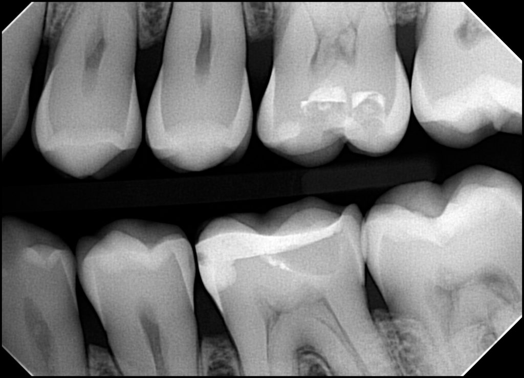
While we have found that different doctors have slightly different preferences when it comes to how they like to see their intraoral radiographs, one common question we get is very simply a preference to make the image lighter or darker.
Sometimes, when the staff member asks about “brightness”, they may actually be referring to “contrast”, which is slightly different. However, for this discussion, we will focus on simple brightness and darkness.
Understanding the Variables
If the staff understands the variables that cause the radiographs to be light or dark, it can empower them to make their own adjustments so as to provide the exact diagnostic information that the doctor is looking for.
To do this, it’s critical that your staff understand a fundamental correlation:
A dental radiograph that is too dark generally means that it was overexposed (i.e. too much radiation hitting the sensor). A dental radiograph that is too light generally means that it was underexposed (i.e. too little radiation hitting the sensor).
The variables that can cause an underexposed or overexposed radiograph span from the size of the patient, the characteristics of the anatomy being captured (e.g. anterior vs posterior), the x-ray parameters and the technique. Of course, this also assumes the equipment is functioning properly.
The obvious question then becomes: what adjustments can the staff make?
Refining Technique
Technique is typically the first area to check if there has been a sudden change in the lightness or darkness of the radiograph (or if this seems to vary based on the operator). For instance, lighter radiographs could be a result of the x-ray source being positioned slightly farther from the sensor. Distance can have a large impact in the amount of radiation hitting the sensor, so positioning the x-ray source slightly farther from the patient’s cheek can result in a lighter radiograph.
Keeping consistent distance can be an even bigger variable if a handheld x-ray sources is used because this device is held “free-hand” [see article about overlooked challenges of handheld].
Adjusting Parameters
If the technique is not the driver, then the operator can look to adjust the parameters of the x-ray sources. The key parameters of most dental x-rays include the kvp (voltage), the mA (current) and the exposure time. Increasing any of these will typically result in a darker radiograph. On most dental x-rays, the parameter that is the easiest to adjust is the exposure time (typically in milliseconds). Increasing the exposure time will typically result in a darker image.
So, therefore, one easy approach is: if a darker radiograph is desired, the staff can increase the exposure time setting on the x-ray. If a lighter radiograph is desired, the staff can decrease the time setting on the x-ray.
Of course, this also assumes the staff is operating x-ray equipment within their specified ranges, and practicing ALARA principles.
Adjusting Filters
As with any digital imaging, software filters can also serve to adjust lightness and darkness. Some common filters that will affect lightness and darkness include: contrast, gamma, or brightness.
It’s also worth noting that some software will auto-adjust the brightness. Sometimes, if the sensor is significantly underexposed for any of the reasons mentioned above, instead of resulting in a light image, it may result in increased graininess. This is due to the software trying to improve the image, when there is less information for the software to work with (due to lack of adequate radiation). It’s a little bit like zooming in on a low resolution image.
Conclusion
Achieving optimal lightness or darkness in dental x-rays requires a comprehensive approach that considers various factors, including technique and equipment settings. By educating the staff on some of these variables it can empower them to be involved in optimizing the images and “course correcting” when problems arise.
Make sure you stay updated on the latest in the dental industry by subscribing