A dental panoramic x-ray machine with cephalometric (sometimes referred to as a dental pan ceph machine) is an essential imaging tool for dentists. This piece of equipment performs two main functions.
First, it operates as a traditional dental panoramic x-ray machine that creates a panoramic radiograph. This is used by the dentist for general exams, as well as third molar, pediatric and extraoral bitewing exams.
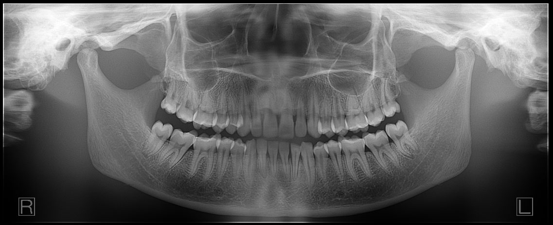
Second, it also allows the dental professional to perform frontal and lateral cephalometric exams. These exams are typically used for orthodontic care. However, they are also used for other evaluations like airway, trauma and overall facial or oral development.
![]() Many dental professionals understand the more obvious questions to ask when purchasing any large piece of equipment, like those having to do with support, training, warranty, etc. However, at ImageWorks, we’ve been providing advanced dental digital x-ray solutions for thousands of offices, and we thought it would be worth sharing some commonly overlooked questions that are really important in selecting the right dental pan ceph x-ray machine.
Many dental professionals understand the more obvious questions to ask when purchasing any large piece of equipment, like those having to do with support, training, warranty, etc. However, at ImageWorks, we’ve been providing advanced dental digital x-ray solutions for thousands of offices, and we thought it would be worth sharing some commonly overlooked questions that are really important in selecting the right dental pan ceph x-ray machine.
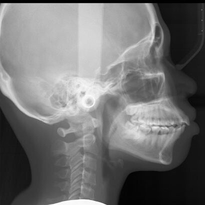
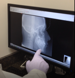
Cephalometric x-ray images are most commonly needed for orthodontic treatment. However, they can also be used for TMJ assessment, sinus evaluation and trauma to the jaw or skull. A cephalometric scan captures both lateral and poster-anterior (PA) images of the entire skull, and these scans are most commonly used as part of orthodontic treatment to measure relative movement of anatomical landmarks.
A somewhat unique characteristic that many dental professionals look for in their cephalometric images is the ability to see soft tissue. Specifically, they use the image to review the relationship a patient’s jaws and teeth have to their soft tissues and the entirety of their skull.
How does the pan ceph x-ray machine differ from a dental pan-only machine or a dental CBCT without ceph?
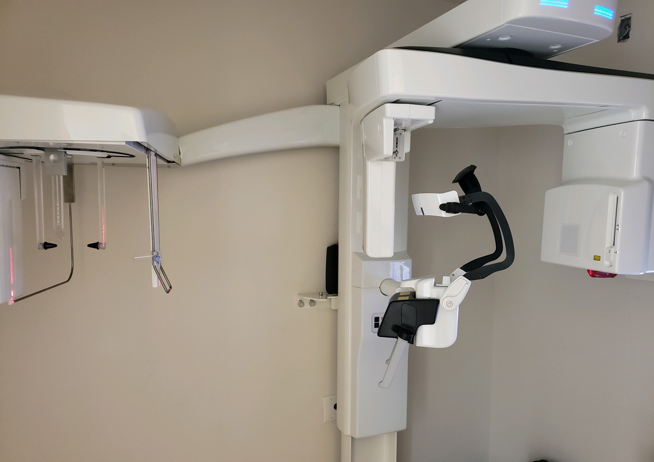
Most cephalometric x-ray machines are combined with traditional dental panoramic x-ray machines, so the machine you purchase will perform both functions. Cephalometric capability can also typically be added to most dental cone beam machines. Typically, the way you can tell that any panoramic x-ray or dental cone beam machine can capture cephalometric x-rays is if the unit has an arm sticking out to the side.
The purpose of this arm is to create distance between the source of the x-ray and the anatomy so that the field of view presented in the image is larger. This is similar to a flashlight that faces a wall. As you move the flashlight away from the wall, the circle of light gets bigger. The patient is positioned at the end of the arm to capture the cephalometric image. The industry standard is that the midsagittal of the patient should be 5 ft from the x-ray source, so the ceph arm will be designed to achieve this.
How good is the pan ceph sensor?
As with dental panoramic x-ray machines, not all systems have the same quality sensors, and the quality of the sensor is a big driver in determining the quality of the images that your machine will produce. Most panoramic and cephalometric sensors today are CMOS design. However, these CMOS sensors typically fall into two basic categories: direct conversion sensors and indirect conversion sensors.
The main difference is that direct conversion sensors use a more expensive material that reduces noise in the signal. As a result, a direct conversion sensor typically is a more expensive component. However, it creates clearer images.
While the enhanced imaging of a direct conversion sensor has an impact with pan-only x-ray machines, it can be more pivotal for a ceph x-ray machine, because soft tissue landmarks are so important for orthodontic applications of cephalometric images.
Will it integrate with my software?
 Currently almost all dental imaging hardware and software is very plug and play when it comes to integration. Almost all dental pan ceph x-ray machines can communicate using the TWAIN standard, and almost all software platforms can accept images from TWAIN devices.
However, one additional configuration that many offices like to have with a dental ceph x-ray machine is that they may have two different software platforms involved. They may have their general imaging software, but they may also utilize orthodontic planning software (sometimes referred to as tracing software). In these situations, they may value a system that can acquire the cephalometric images directly into their tracing program, while acquiring their panoramic images into their general imaging software.
Does the pan ceph equipment have one sensor or two?
In order for a dental ceph x-ray machine to also perform panoramic scans, the machine must be able to capture images in two different locations: the panoramic scan is typically done when the patient is standing near the column (close to the x-ray source), and the cephalometric scan when the patient is standing at the end of the cephalometric arm (farther from the x-ray source).
 In each of these scenarios, a sensor must be located near the patient to capture the image. This creates a challenge for a manufacturer, because typically the sensor is at the heart of the quality of the system, and therefore is one of the more expensive components – so having two sensors can make the system cost too high.
So dental manufacturers typically solve this problem in one of two ways: either the equipment has a lower-resolution, cheaper sensor in both the panoramic image location and the cephalometric image location (two sensors), or it has a single higher-resolution sensor that can be moved between the different positions. As technology has advanced, the mechanisms in single sensor platforms to move the sensor have become so easy to use and mistake-proof, this has become a common design to get the best of both worlds: maximizing image quality while still being easy to use.
Are ceph images captured with a scan or with a single exposure?
Most dental cephalometric x-ray machines capture the cephalometric image by performing a quick scan: usually 6-10 seconds. However, one variation of the two-sensor design mentioned above is to utilize a larger “flat-panel” style sensor at the end of the arm for cephalometric images. The main advantage of this style is that instead of a scan, the cephalometric image is captured with a single exposure.
We hope you find this useful. If you have any questions or would like to speak with a specialist to find the right fit for your practice, click below. We would love to talk to you!
Subscribe to Receive More Great Articles