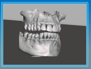
As more and more of the world of dentistry moves into 3D, there is a collision of two different data standards: DICOM and STL. To understand these standards and the roles that they play, it may be helpful to understand where they come from.
The first data format is called DICOM, which stands for Digital Imaging and Communications in Medicine. For decades, this has been the standard for all medical digital radiography. It not only covers the formats to be used for storage of digital medical images, but also covers the protocols related to the communication services which are useful in the medical imaging workflow. Now, in the dental arena, digital radiographs are typically DICOM standard mainly to drive consistency in image file format. The protocol standards that are defined by DICOM are less relevant to a typical dental office because they are used when communicating with a Picture Archiving and Communication System (PACS). PACS are used in larger facilities (e.g. hospitals, prisons) that have a wide variety of digital radiographs that need to be performed, stored and managed.
In a dental office, the traditional cone beam scan are typically stored by the imaging software in DICOM format. For a dental cone beam, the data that makes up the volumes actually consist of many (typically hundreds) of 2D slices. Each of these slices is also typically in DICOM format.

The second data format that has become more important to dentistry recently is STL, which is short for stereolithography (You might also hear it referred to as Standard Triangle Language or Standard Tessellation Language). This format has it’s origins in the fields of 3D printing and Computer Aided Design and Computer Aided Manufacturing (sometimes referred to as CAD/CAM). It describes the surface geometry of a three-dimensional object and has become the data format that most 3D printers and milling systems require. In a dental office, the traditional intraoral scanner will output in STL format.
While the DICOM approach to 3D breaks the volume into slices. The STL format breaks the surface of the volume down into “tiles” which are typically triangular. As a result, the DICOM file tends to provide more information about what’s inside the volume, while the STL file tends to provide more information about the surface of the volume.
When creating an implant plan, clearly both of these types of information become very useful. Particularly when a surgical guide is being created because it utilizes information underneath the surface but also needs to know information about the surface itself including the soft tissue.
Commonly, the dental office does not need to worry about converting one format to the other because this is generally where the lab plays a role. The lab has deep expertise and software tools to help them merge these different formats together to create the implant plan for the dental professional. However, if a dental office would like to start performing chairside milling, or printing their own surgical guides, then it becomes more important to understand how these data formats work together.
