
University of Texas School of Dentistry to Implement Next Generation Dental Microscope

Dr. David Cochran, Dr. Angela Palaiologou-Gallis and Dr. Motofumi Yamashita

University of Texas School of Dentistry to Implement Next Generation Dental Microscope


The Department of Periodontics at UT Health San Antonio School of Dentistry has implemented the next generation Mielscope dental microscope as part of its continuing drive to train the next generation of dental professionals on the latest in cutting edge tools. 

The Mielscope Microscope, implemented by Imageworks based on technology developed by Yoshida, enables the doctor to get the magnification, clarity and ergonomics of a traditional dental microscope without the steep learning curve and limited application of a traditional microscope.

During the event, Dr. David Cochran, the chairman of the Department of Periodontics shared his view of how the Mielscope will help empower new dentists to care for their patients. 

Dr. Motofumi Yamashita, who has been using the Mielscope in his practice, then presented a series of powerful cases showing how the Mielscope helps his practice run more quickly and precisely.  He presented stunning videos of a myriad examples including a crack identification, tooth preparation, endodontics, periodontal surgery, PDL visibility, bone grafts, papilla preservation, ridge preservation, sinus lifts and much more.

He showed that many traditional microscopes have a shallow depth of focus, which can result in only a small portion of the field of view being in focus.  Conversely, the Mielscope’s large depth of focus means the anatomy across the entire screen presents in sharp focus.

Dr. Yamashita then discussed the eye fatigue that can arise from extended periods of looking through binocular eyepieces or loupes, and explained how the Mielscope’s screen allows the operator to avoid this fatigue.  He further emphasized that avoiding this fatigue means the doctor can better focus on the patient.  Eliminating the eyepieces also means there is almost no learning curve as the Mielscope is intuitive and simple to use.

He explained how the high-precision articulating arm of the Mielscope allows the doctor to visualize anatomy that would be very difficult to see with a traditional microscope.  In addition, he showed how the high magnification and 4K imagery across large fields of view assists him with laser procedures and implant procedures.

He also showed how the fantastic imagery can be used to assist in patient communication and can make assisted procedures run faster and more efficiently because the entire team can see exactly what the doctors sees.

Finally, Dr. Yamashita showed how the Mielscope has even enhanced the profitability of his hygiene program, as it allows the hygienist to perform more effectively and more quickly.  In addition, the fantastic imagery allows high resolution “before” and “after” images which presents compelling evidence to the patient that they should keep their cleaning appointments.
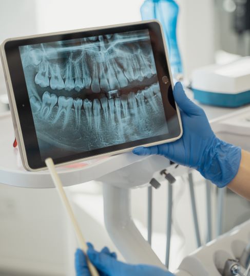
 

Learn more about the Mielscope

Dr. Boggess Shares a Unique Case From His Pan Ceph System


Dr. Vaughn Boggess is an orthodontist in Lexington, Kentucky.  His pan ceph went down and put him in a bind because he needed to get up and running fast, so he wanted a great solution that his staff would easily be able to use.  He shares his experience getting back online with the Imageworks Pan Ceph system, and shares an interesting pedo case. 


Learn more about the Panoura X-era from ImageWorks


Subscribe to Receive More Great Articles

    Medical Applications of Dental Panoramic X-Rays in Cardiac Surgery

    Dental panoramic X-rays play a crucial role in the medical management of patients undergoing cardiac valve surgery. These radiographic images can help surgeons identify and treat issues that may complicate cardiac procedures. This article explores the importance of dental panoramic X-rays in the context of cardiac surgery, highlighting their role in preventing complications and ensuring optimal patient outcomes.
     

    The Importance of Dental Health in Cardiac Surgery

    Cardiac valve surgery, which includes procedures on both native and prosthetic valves, increases the risk of postoperative Infective Endocarditis (IE).1  IE is an infection of the inner lining of the heart chambers and valves, often caused by bacteria entering the bloodstream from oral infections. Research indicates that approximately 10-20% of IE cases are linked to oral infections, with chronic periodontitis significantly raising the risk, especially in patients with pre-existing heart conditions.


    Preoperative Dental Evaluation

    Patients scheduled for cardiac valve surgery should undergo a comprehensive dental evaluation before the procedure. This evaluation includes a detailed medical history review, an examination of medications and allergies, and a thorough oral and dental examination. Panoramic X-rays are essential in this process, providing a broad view of the oral cavity that helps identify any potential sources of infection.

    The initial dental assessment aims to detect and address oral infectious foci, such as caries, periodontal disease, and endodontic issues. By treating these conditions preoperatively, the risk of bacteremia—and consequently IE—during and after cardiac surgery is significantly reduced.


    Dental Panoramic X-Rays: A Comprehensive Tool

    Panoramic X-rays are particularly valuable in the dental management of cardiac surgery patients for several reasons:


    1. Broad Coverage: These X-rays capture the entire mouth in a single image, including the teeth, jaws, and surrounding structures. This wide coverage is crucial for identifying hidden issues that might not be visible in a standard dental examination.
    2. Early Detection: Panoramic X-rays can reveal early signs of periodontal disease, caries, and other dental problems. Early detection allows for timely intervention, reducing the risk of complications during cardiac surgery.
    3. Surgical Planning: For patients requiring extractions or other dental procedures, panoramic X-rays provide detailed information that aids in surgical planning. They help determine the extent of dental issues and the best approach for treatment, ensuring minimal disruption to the patient’s overall health.


    Antibiotic Prophylaxis and Anticoagulant Management

    Patients who have had valve surgery have a high risk of IE, and therefore may require antibiotic prophylaxis before dental procedures. The American Heart Association and the American College of Cardiology recommend this for patients with a history of IE, prosthetic heart valves, congenital heart disease, and heart transplants with valvular dysfunction. The antibiotics are typically administered 30-60 minutes before dental procedures that are likely to cause bacteremia, such as extractions and periodontal treatments.


    Conclusion

    Dental panoramic X-rays are indispensable in the medical management of patients undergoing cardiac valve surgery. They provide a comprehensive view of the oral cavity, enabling early detection and treatment of dental issues that could lead to serious complications like Infective Endocarditis. By integrating these radiographic assessments into the preoperative planning for cardiac surgery patients, healthcare providers can significantly enhance patient safety and surgical outcomes. Ensuring optimal dental health through panoramic X-rays and appropriate prophylactic measures is a critical step in the holistic care of cardiac surgery patients.


    1. Souza AF, Rocha AL, Castro WH, Gelape CL, Nunes MCP, Oliveira SR, Travassos DV, Silva TA. Dental management for patients undergoing heart valve surgery. J Card Surg. 2017 Oct;32(10):627-632. doi: 10.1111/jocs.13211. Epub 2017 Sep 12. PMID: 28898929.

    Make sure you stay updated on the latest in the dental industry by subscribing

      8 Reasons Your Dental Panoramic Radiographs Look Horrible (and How To Avoid Them)

      This is a training module that we provide to all of our offices using ImageWorks cone beam and panoramic modalities.  Dental professionals have told us it’s been very useful to help heir staff make he most of their investments.  We wanted to share with our community, because the lessons hold true regardless of type of equipment you are using.  


      Make sure you stay updated on the latest in the dental industry by subscribing


        Dental Cone Beam and Panoramic CDT Codes


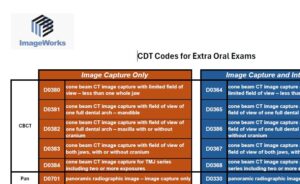
        We are often asked for the applicable CDT codes for Cone Beam and Panoramic Radiographs, so we wanted to share a resources that summarizes these codes for the most commonly used extraoral exams. 

        Current Dental Terminology (CDT) codes are alphanumeric codes used to document dental procedures and report them to dental payers. The American Dental Association (ADA) developed and maintains the CDT codes as a standardized language to help dentists and other healthcare professionals communicate

        You can also download the resource by clicking on the image.

        Click to open


        Make sure you stay updated on the latest in the dental industry by subscribing


          Viewing Calcification of Carotid Arteries in a Panoramic Radiograph


          An interesting topic came up in an online dental forum around what to do when the dental professional notices an indication on a panoramic radiograph that resembles calcification in the carotid artery. 

          Based on this, we wanted to provide a few resources that may be useful for dental staff to be aware of this indication along with potential implications.


          Make sure you stay updated on the latest in the dental industry by subscribing


            Carotid artery calcifications (CAC) are hard, calcified deposits that form in the carotid arteries, which are the major blood vessels in the neck. There are two carotid arteries, one on each side of the neck: the right and left common carotid arteries. Each common carotid artery branches into the internal carotid artery, which supplies blood to the brain, and the external carotid artery, which supplies blood to the face and neck.

            These calcifications can indicate a higher risk of cardiovascular diseases like stroke and heart attack. Detecting these early can be vital for preventing serious health issues.

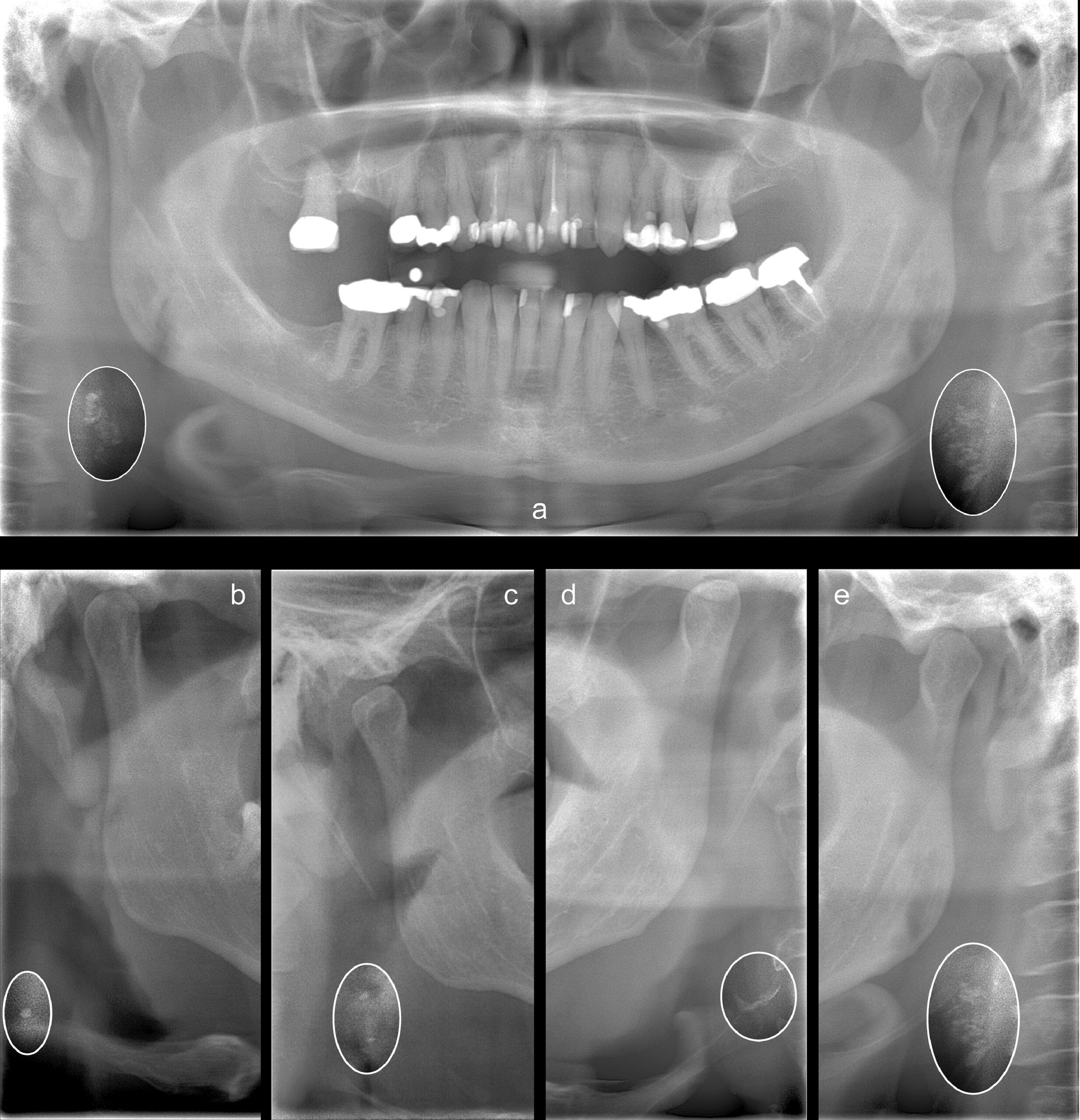
            Dental panoramic radiographs can also show the carotid arteries. When dentists review these radiographs, they might notice radiopaque nodular lesions, which can indicate CAC. According to Friedlander et al. [1], these lesions appear separate from the hyoid bone and are adjacent to the cervical vertebrae, usually between the C3 and C4 vertebrae or below. This is close to the location where the carotid artery bifurcates.


            Research Findings

            Studies such as Magnus Bladh et. al [2]  have shown a strong correlation between the presence of CAC on panoramic radiographs and more advanced carotid atherosclerosis detected by ultrasound (US). Ultrasound is a well-established method for detecting soft tissue changes and plaques in the arteries, which are significant markers for cardiovascular disease. The findings suggest that if CAC is detected on a panoramic radiograph, there is a high probability that more severe atherosclerotic changes will be found upon further examination with ultrasound.

            Here are a few sample radiographs from this study:


            Understanding the panoramic x-ray projection

            Because all panoramic radiographs are projections, it’s important to be aware that the farther from the focal trough the anatomy is, the more distorted it may appear.  In this situation, distortion is not as important because it’s more a matter of flagging the indication.  However, if you are using a panoramic x-ray machine that has the ability to capture multiple focal troughs, then it is possible to gather additional views from the single scan.  In other words, these panoramic x-ray machines allow the operator to extract more information from the scan by evaluating focal planes that may not be as ideal for the dentition, but more ideal to view the carotid artery.


            Advantages and Limitations

            The ability to detect CAC on panoramic radiographs can be particularly useful in both general and specialized dentistry. Dentists who identify these calcifications can recommend that patients seek medical care for further evaluation and preventive treatment. This can help reduce the risk of CVD-related issues.

            However, it’s important to note that not all carotid plaques are calcified, and sometimes the plaques may be located outside the area captured by the X-ray. Therefore, a negative finding on a panoramic radiograph does not necessarily mean that the patient has no vascular issues.

            Despite some limitations, the high specificity of CAC detected on panoramic radiographs, coupled with their correlation to ultrasound findings, underscores their importance. While ultrasound can visualize the soft tissue components of plaques that X-rays cannot, panoramic X-rays excel at detecting even small calcifications [2].

            In some cases, calcifications may not be visible on panoramic radiographs if they are situated below the imaging area. Yet, when CACs are detected, particularly when they are bilateral and outline the vessel, it strongly suggests significant vascular changes that require medical attention [2].


            Conclusion

            In summary, panoramic radiographs are a valuable tool in dental practice not only for assessing dental health but also for identifying potential cardiovascular risks. Dentists who recognize CACs on these radiographs can play a crucial role in early detection and prevention of cardiovascular disease. By recommending further medical evaluation for patients with detected CACs, dentists can contribute to improved cardiovascular health outcomes.

            While panoramic X-rays should not replace traditional cardiovascular screenings, they offer an additional layer of detection that can be lifesaving. For patients, this means that routine dental exams could also provide important insights into their overall health, highlighting the interconnected nature of dental and medical care.

            1.  AH Friedlander, NR Garrett, EE Chin, JD. Baker; Ultrasonographic confirmation of carotid artery atheromas diagnosed via panoramic radiography; J Am Dent Assoc, 136 (2005), pp. 635-640
            2.  Magnus Bladh, DDS,a Nils Gustafsson, DDS, PhD, et.al; Defined shapes of carotid artery calcifications on panoramic radiographs correlate with specific signs of cardiovascular disease on ultrasound examination; Oral Surgery, Oral Medicine, Oral Pathology and Oral Radiology Volume 137, Issue 4, April 2024, Pages 408-420


            Make sure you stay updated on the latest in the dental industry by subscribing


              Dental Hygienist and Dental Assistant Wage Analysis

              One of the biggest challenges that dentists and dental organizations face is staff turnover. This has been especially acute during the upheaval in a post-COVID inflationary world.  Having great staff is paramount in operating an efficient practice that provides superior patient care.  However, overpaying for staff can mean other parts of the practice get short changed.

              To the help the dental practice solve this problem of optimizing, the most powerful tool is data.  However, overly broad and general information isn’t really all that helpful – because every economy is local. 


              Make sure you stay updated on the latest in the dental industry by subscribing


                Given this, ImageWorks has done an in-depth analysis based the latest dental employee wage data from the Bureau of Labor and Statistics.  This data was released in April of 2024.

                The report below includes data on median and upper percentile wage data for dental hygienists and dental assistants across over 500 regions in the US.  This data is useful to help dentists and practice owners audit of their own employment incentives because you can find data specific to your region.

                Based on BLS data, you can see how he rate of inflation affected the national median wage for dental assistants and hygienists.  While assistants saw a more dramatic change year over year, it is starting to normalize.  However, hygienists have seen steadier growth that has persisted into the most recent year recorded. 

                Download the in depth regional report below to see how your employees compare in your region.


                Download Hygienist and Dental Assistant Regional Wage Report


                Make sure you stay updated on the latest in the dental industry by subscribing


                  Three Common Pitfalls When Moving a Dental CBCT Machine

                  As dental offices grow, restructure, and acquire new offices, ownership is commonly in a situation where they need to move assets and equipment from one location to another.  For many dental offices, the most valuable piece of equipment is their dental cone beam ct system

                  As a result, many dental offices are interested in physically moving that dental cone beam system to a different location from where it was installed.  However, moving this kind of equipment can be tricky not only because it is a valuable asset, but also because there are a number of issues that can cause problems with the equipment after move.  Ironically, most of these items are very easily avoided if the owner or the mover is aware of them.


                  Recalibration or realignment may be needed

                  All dental cone beam systems have a rotating chassis that has a sensor on one side and an x-ray source on the other.  In order to take high resolution images, these two pieces have to be precisely aligned with each other and with the patient positioning mechanism of the machine. 

                  If the rotational assembly is detached from the main column of the unit, this will typically offset the alignment of the unit such that the unit will need to be realigned in the new location.  Moving the unit without any disassembly may still require recalibration or realignment in the new location as abnormal vibration or movement of the components may also cause the unit to fall out of alignment.


                  Internal components may break

                  A dental cone beam machine is not typically designed to be transported in its fully-assembled configuration.  While a good partner performing the move will support and protect the equipment to avoid obvious damage, it’s worth noting that sometimes damage to internal components can be done during the move that may not be outwardly obvious. 

                  One common area of vulnerability for many units is inside the c-arm assembly.  For example, many dental cone beams have highly sensitive gears, motors, sensors, and potentiometers that are not designed to withstand undue force or stress that may come from vibration or fast movement that may come with rough transport. Furthermore, failure of these mechanisms may not be obvious when the unit is installed in the new location.

                  To reduce this risk, most experienced technicians will fully bind the unit up (e.g. with shrink wrap) before it is moved.  However, in addition, be aware of how the assembly may move when it rests in different positions.  For instance, even though the unit is fully shrink wrapped in its upright position, there may be unexpected degrees of freedom when the unit is laid on it’s back. Gently push and pull on different areas of the c-arm when it is in its “transport position” to assure there’s no ability to shift or move during transport.


                  Changes to approved shielding study

                  If your office is located in a state (or locality) that requires shielding plan approval, then moving the unit to a new location (or even a different position in an existing office) may mean that the original shielding study is no longer valid. 

                  Some common characteristics that shielding studies take into account are not only the obvious things like the material of the walls and the layout of the room, but also what activity is occurring adjacent to the unit – not only on the other side of the wall, but also above and below. 

                  If the assumptions of the initial review are not the same, it’s possible that an inspector will identify that the shielding study is no longer valid.


                  Conclusion

                  If you are planning a move for your dental cone beam, simply planning for these items will often reduce the probability of any surprises when you look to go live in your new location


                  Make sure you stay updated on the latest in the dental industry by subscribing


                    How to Solve One of the Biggest Challenges Dentists Have With Microscopes

                    How dentists overcome one of the biggest challenges they have with dental microscopes


                    Learn more about the Mielscope microscope from ImageWorks


                    Biggest Challenge Dentists Have with Dental Microscopes2

                    How dentists overcome one of the biggest challenges they have with dental microscopes


                    Learn more about the Mielscope microscope from ImageWorks