
Cephalometric radiographs are most commonly needed for orthodontic treatment. However, they can also be used for TMJ assessment, sinus evaluation and trauma to the jaw or skull. A cephalometric scan captures both lateral and poster-anterior (PA) images of the entire skull, and these scans are most commonly used as part of orthodontic treatment to measure relative movement of anatomical landmarks.
Important Overlooked Questions When Buying a Dental Pan Ceph or Cone Beam With Ceph X-Ray Machine
Typically, the way you can tell that any panoramic x-ray or dental cone beam machine can capture cephalometric x-rays is if the unit has an arm sticking out to the side.
The purpose of this arm is to create distance between the source of the x-ray and the anatomy so that the field of view presented in the image is larger. This is similar to a flashlight that faces a wall. As you move the flashlight away from the wall, the circle of light gets bigger. The patient is positioned at the end of the arm to capture the cephalometric image. The industry standard is that the midsagittal of the patient should be 5 ft from the x-ray source, so the ceph arm will be designed to achieve this.
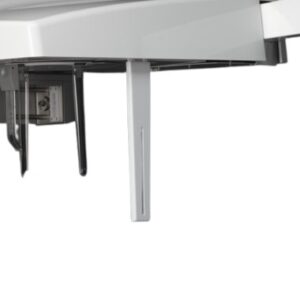 A small but really important component of the the pan ceph is called the secondary collimator. A collimator is a general term for an opening that only allows x-ray through that is going to hit the sensor, and therefore play a role in creating the desired radiograph. Any x-ray energy that would not hit this sensor is blocked by the collimator, and therefore shielded from the patient. All dental x-ray machines have a collimator.
However, with a dental ceph machine, because the patient stands farther away from the source of the x-ray (at the end of the ceph arm), there is typically another collimator found at the end of the arm. It will typically look like a vertical flange with a slit down the middle, and it will sit between the patient and the source of the x-ray.
Typically, a key part of aligning the ceph involves making sure that the primary collimator (near the source of the x-ray) is “aligned” with the secondary collimator at the end of the ceph arm. A poorly aligned secondary collimator can affect the resulting image because it may be blocking some of the required x-ray that the ceph sensor requires to take a good ceph radiograph.

Learn more about Cone Beam Solutions
Learn more about Panoramic and Pan Ceph Solutions